01
What to know
- The glans is the sensitive head; the shaft contains erectile tissue that fills with blood during an erection.
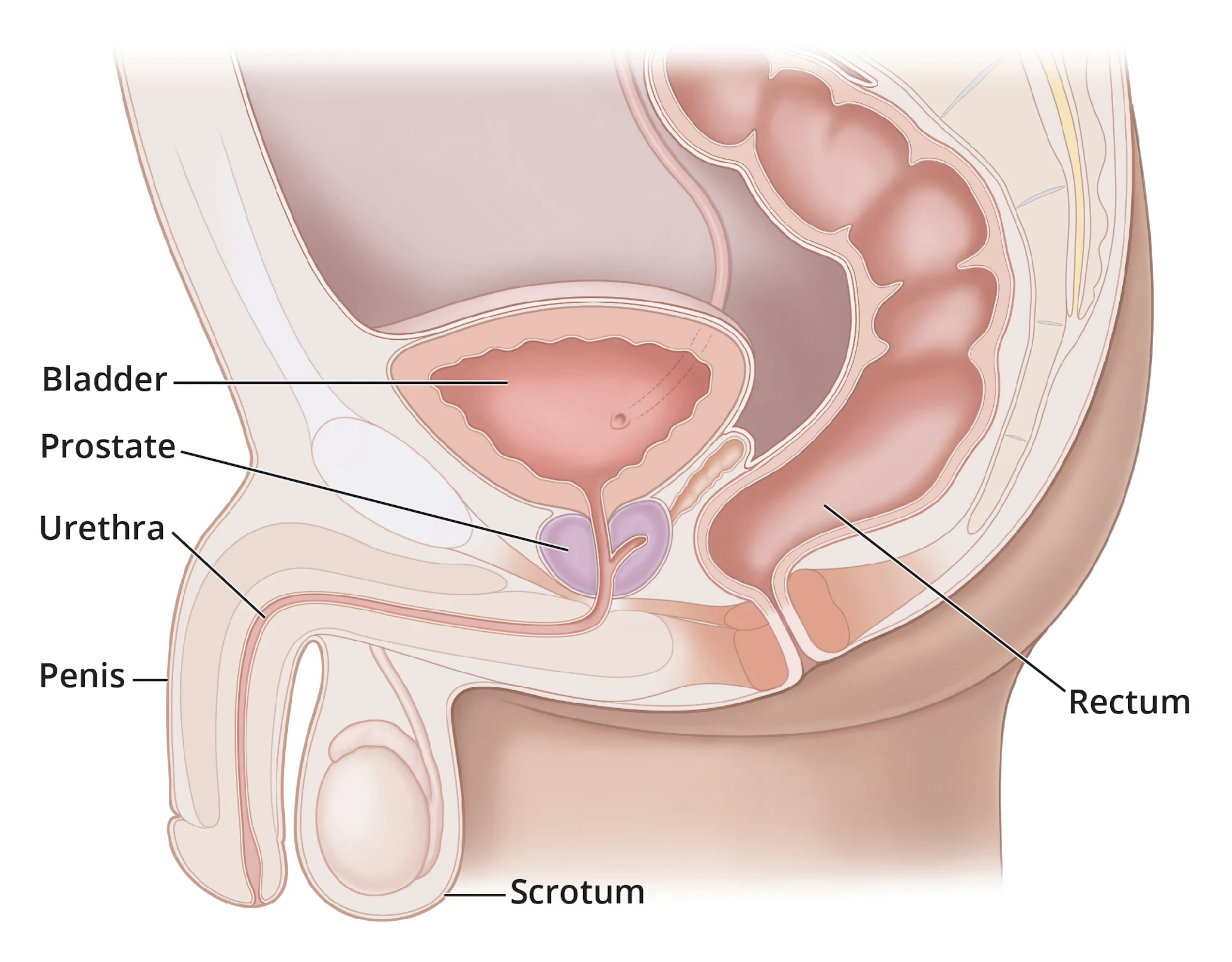
- The urethra carries urine and semen at different times. Burning, discharge, blood, or inability to urinate is not simply an appearance question.
- Foreskin mobility varies with age and anatomy. It should never be forced, especially in children.
- Sensation, angle, veins, skin tone, and resting size vary between people and can change with temperature, stress, medication, and age.
What people often get wrong
A similar-looking photo or one matching symptom cannot confirm a cause. Timing, change, examination, and sometimes testing matter.
02
Useful next steps
- Learn your usual baseline without repeatedly checking or comparing.
- Describe a change by location, timing, pain, and whether it affects urination or sexual function.
When to get care
Do not rely on guesswork when the pattern changes.
- Seek care for a new persistent lump, sore, discharge, bleeding, major color change, pain, or loss of function.
03
What a clinician may ask
- When did you first notice it, and is it changing?
- Is there pain, discharge, bleeding, fever, urinary change, or loss of function?
- Were there recent medicines, illness, injury, sexual contact, products, or procedures?